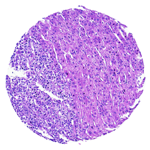
Liver Tumour Tissue Microarray
Liver tumour tissue microarray (TMA) is an advanced platform designed to accelerate high-throughput studies of liver cancer by allowing multiple tissue samples to be examined on a single slide. The process involves carefully extracting small core biopsies from various liver tumour specimens and embedding them into one paraffin block. This consolidated format enables researchers to investigate biomarker expression, tumour heterogeneity, pathways of disease progression, and potential therapeutic targets across a broad range of cases with consistency and efficiency. By standardizing sample comparison, liver tumour TMA provides a reliable foundation for translational research and the development of targeted treatments.
Our Liver Tumour Tissue Microarrays
Our liver tumour tissue microarray is designed to streamline cancer research by enabling high-throughput analysis of tissue samples. TMAs are particularly valuable for studying liver cancer, allowing efficient examination of biomarkers, molecular profiles, and pathological characteristics. With the ArrayZeal platform, MYmAb Biologics supplies liver tumour tissue microarrays sourced from Southeast Asian populations. Obtain a liver TMA from MYmAb Biologics to accelerate your cancer research and support more comprehensive bladder cancer studies for the global population.
Benefits of Liver Tumour Tissue Microarrays
Liver tumour tissue microarrays offer numerous advantages for researchers seeking efficient, large-scale tissue analysis:
- Efficiency: Evaluate hundreds of samples on a single platform, streamlining workflows.
- Cost-Effectiveness: Consolidated sample testing lowers laboratory expenses and reagent usage.
- Reproducibility: Uniformly prepared TMAs ensure reproducible and reliable outcomes across experiments.
- Comprehensive Data: Analyse diverse tumour subtypes and stages in one compact format.
- Customisability: Custom TMAs tailored to specific research questions and goals.
Applications of Liver Tumour TMAs in Cancer Research
- Biomarker Discovery: Identify and validate biomarkers for liver cancer diagnosis, prognosis, and treatment.
- Drug Development: Evaluate drug efficacy and predict therapeutic responses using diverse tumour samples.
- Histological Studies: Explore tumour morphology and progression across a wide spectrum of cases.
- Genomic and Proteomic Analysis: Perform high-throughput molecular studies to uncover genetic and protein-level changes in liver cancer.
- Precision Medicine Research: Support research efforts toward tailored cancer therapies based on individual patient profiles.
Why Choose Our Liver Tumour Tissue Microarray
MYmAb Biologics ethically produces human tissue microarrays (TMAs) through our ArrayZeal platform, guided by strict ethical standards and built on a foundation of precision and reliability. Each TMA is carefully constructed using clinically validated human tissue samples sourced from diverse Southeast Asian populations. This ensure better representation of ethic genetic variability, which is an important factor that influences disease progression and treatment responses. Committed to delivering reliable and reproducible results, ArrayZeal support robust scientific discoveries and accelerate translational research. By offering Southeast Asian-derived TMAs, we empower researchers to explore novel therapies within a broader genetic context, paving the way for groundbreaking discoveries and personalized medicine.
Frequently Asked Questions
Conventionally, immunohistochemical staining has been performed on whole tissue sections. However, this traditional method, which requires the processing and staining of hundreds or even thousands of slides, is time-consuming and expensive. In contrast, a TMA allows simultaneous analysis of hundreds of tissues in one go using identical conditions. Thus, TMA technology markedly conserves reagents, saves time and greatly decreases the amount of archival tissue required for a particular study, thus preserving the tissues for other diagnostic and/or research needs.
A core size of 0.6 to 2.0 mm is adequate to represent the whole tissue section.
Sometimes, a small core of sample may not be representative of the whole tumour, especially in heterogenous tumour types. To overcome this, we offer TMAs that include two to three cores of samples from different locations of a single tumour block.
Negative and positive control TMA slides should be included as they are important in ensuring the validity of the experiment.
To help with TMA orientation.
A minimum of 3 slides per TMA type to include positive and negative controls.
All patients are anonymised.
Yes. You may contact us for further details and discuss on your needs.
The TMAs are sent in ambient temperature.
We recommend you to store your TMA slides at 4°C for not more than a year.
Request for Quote
Location:
Block D-G-8, UPM-MTDC Technology Centre III, Universiti Putra Malaysia, 43400 Serdang, Selangor Darul Ehsan, Malaysia.
Phone:
03-8938 9819
Email:
info.mymab@gmail.com

