Tissue Microarray (TMA): The Future of Tissue-Based Research and Diagnostics
In modern biomedical research, efficiency and precision define progress. One of the most impactful technologies driving that progress is the tissue microarray (TMA) — a platform that allows researchers to analyze hundreds of tissue samples simultaneously under identical conditions. For laboratories, hospitals, and biotech companies aiming to accelerate biomarker discovery or streamline histopathological analysis, tissue microarrays are transforming how tissue-based studies are conducted.
At MYmAb Biologics, we specialize in the design, fabrication, and optimization of high-quality tissue microarrays that empower global researchers and diagnostics companies to conduct large-scale, reproducible studies with greater efficiency and clarity.
What is a Tissue Microarray (TMA)?
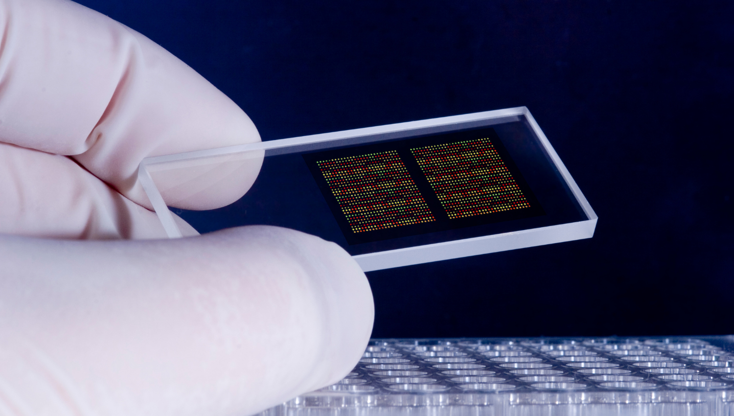
A tissue microarray is a high-throughput platform that consolidates small core biopsies from multiple tissue samples into a single paraffin block. Each block can contain anywhere from dozens to thousands of distinct tissue cores arranged in a defined grid. Once sectioned and mounted on slides, this block enables simultaneous analysis — whether for immunohistochemistry (IHC), in-situ hybridization, or molecular marker validation.
The concept is simple yet powerful: instead of analyzing one sample at a time, a TMA allows researchers to analyze hundreds of samples under the same experimental conditions, ensuring consistency and saving substantial time and resources.
At MYmAb Biologics, we ensure that every tissue microarray is meticulously designed for accuracy, using optimized paraffin quality, precision coring tools, and validated workflows that align with both clinical and research-grade standards.
How Tissue Microarrays Are Made
Creating a tissue microarray involves a series of controlled, high-precision steps. Each phase impacts the reproducibility and quality of the final analysis.
- Donor Block Selection
- Tissue Coring
- Recipient Block Construction
- Sectioning and Staining
1. Donor Block Selection
Experienced pathologists identify regions of interest on formalin-fixed paraffin-embedded (FFPE) donor blocks. These could represent tumor regions, normal tissues, or disease-specific areas depending on the study objective.
2. Tissue Coring
Using a specialized punching device, small cylindrical cores (typically 0.6–2.0 mm in diameter) are extracted from the donor blocks. At MYmAb Biologics, we employ high-precision core punches that maintain tissue integrity and avoid distortion — a crucial factor for downstream staining quality.
3. Recipient Block Construction
The tissue cores are inserted into a recipient paraffin block, pre-marked with coordinates that maintain accurate sample tracking. Each block is carefully embedded with low-melting paraffin to ensure stability during sectioning.
4. Sectioning and Staining
Once the array is set, the block is sectioned into thin slices (usually 3–5 µm thick) and mounted onto glass slides. These slides are then stained or processed for various molecular assays — including immunofluorescence, IHC, and nucleic acid hybridization — depending on research needs.
This process allows researchers to analyze hundreds of tissue cores side by side, reducing reagent consumption and experimental variability.
Why Tissue Microarrays Matter
Tissue microarrays bridge the gap between traditional pathology and high-throughput molecular biology. They deliver several key advantages:
- High-Throughput Analysis
- Consistency and Reproducibility
- Cost and Resource Efficiency
- Preservation of Valuable Time
1. High-Throughput Analysis
A single TMA can contain up to thousands of tissue cores, allowing simultaneous analysis across large patient cohorts. This scalability is invaluable in oncology, where biomarker validation requires testing on diverse tumor samples.
2. Consistency and Reproducibility
By ensuring all tissues are processed under identical conditions, TMAs minimize technical variability — enabling consistent comparison across samples, batches, or laboratories.
3. Cost and Resource Efficiency
TMAs significantly reduce the use of reagents, antibodies, and technician hours. For research facilities managing limited budgets or rare tissue archives, this efficiency can translate to exponential cost savings.
4. Preservation of Valuable Tissue
Because each core uses only a small section of the original specimen, TMAs allow researchers to maximize data extraction from scarce or irreplaceable tissues — a critical advantage in studies involving rare diseases or archival samples.
At MYmAb Biologics, our custom TMAs are engineered to optimize these benefits, providing research institutions and diagnostics companies with standardized, reproducible, and cost-efficient tissue analysis platforms.
Applications of Tissue Microarrays
Tissue microarrays have evolved into indispensable tools across multiple disciplines of biomedical science and clinical diagnostics.
- Oncology Research
- Drug Discovery and Development
- Clinical Diagnostics and Personalized Medicine
- Academic and Translational Research
1. Oncology Research
The most widespread application of TMAs is in cancer research. They enable large-scale analysis of tumor biomarkers, helping identify molecular signatures linked to disease progression, prognosis, or therapeutic response. At MYmAb Biologics, we collaborate with oncological research teams to produce specialized tumor microarrays that support drug development and biomarker validation.
2. Drug Discovery and Development
Pharmaceutical companies utilize TMAs to screen drug targets and assess biomarker expression in preclinical studies. By analyzing hundreds of samples on a single slide, researchers can efficiently test how candidate compounds interact with specific cellular markers, accelerating the pipeline from discovery to clinical validation.
3. Clinical Diagnostics and Personalized Medicine
TMAs have become integral to developing diagnostic assays that correlate molecular data with patient outcomes. They support the validation of predictive markers, contributing to the advancement of personalized medicine — tailoring treatments to the unique molecular profiles of patients.
4. Academic and Translational Research
Universities and research institutes rely on TMAs for molecular pathology, comparative genomics, and proteomics. The reproducibility and scalability of arrays make them ideal for hypothesis testing and longitudinal studies.
MYmAb Biologics supports global research projects by providing custom-designed TMAs built to match specific tissue types, study designs, and biomarker targets.
Limitations and Quality Considerations
While tissue microarrays offer substantial advantages, challenges such as tissue heterogeneity can affect data interpretation. Since each core represents a small fraction of the original tissue, variations within tumors may not be fully captured.
At MYmAb Biologics, we mitigate this limitation through strategic multi-core sampling, advanced imaging validation, and stringent quality control protocols. This ensures that every TMA block we produce maintains the highest possible representativeness and reliability.
The Future of Tissue Microarrays
The next evolution of TMAs lies in automation and digital transformation. Automated tissue arrayers are now capable of coring and mapping thousands of samples with robotic precision. Meanwhile, digital pathology platforms enable slides to be scanned, stored, and analyzed using AI algorithms — enhancing pattern recognition and quantitative analysis.
MYmAb Biologics is actively integrating these innovations, combining automated array construction, AI-assisted image analytics, and cloud-based data integration to deliver the next generation of tissue microarray technology. Our goal is to make tissue-based analysis faster, more accurate, and universally accessible to researchers worldwide.
Why Choose MYmAb Biologics
At MYmAb Biologics, we are more than just a tissue microarray manufacturer — we are a partner in advancing tissue-based discovery. Our team combines technical expertise with innovation to help research institutions, hospitals, and biotech firms achieve reproducible, scalable results.
We offer:
Custom tissue microarray design and construction
Human and animal model tissue options
Automated core mapping and labeling
Quality assurance and histological validation
Global shipping and collaboration support
Whether your focus is oncology, pathology, or translational research, MYmAb Biologics provides the reliability, precision, and scalability you need to push discovery forward.
Final Thoughts
Tissue microarrays represent the intersection of efficiency, precision, and innovation in tissue-based research. As global demand for scalable molecular diagnostics grows, TMAs will remain a cornerstone in biomarker discovery and personalized medicine.
At MYmAb Biologics, we are committed to advancing this frontier — delivering the quality, accuracy, and innovation researchers need to accelerate the future of medical science.